| Leptospirosis | ||
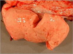
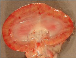
Weil’s syndrome, Swamp fever, Mud fever, Autumn fever (Akiyami), Swineherd’s disease, Rice-field fever, Cane-cutter’s fever, Hemorrhagic jaundice, Stuttgart disease, Canicola fever, Redwater of calves. Etiology Leptospirosis is caused by various species of Leptospira, a spirochete in the family Leptospiraceae, order Spirochaetales. The classification of this organism is complex. Before 1989, all of the pathogenic isolates belonged to the species Leptospira interrogans, which contained more than 200 serovars in 23 serogroups. More recently, the genus Leptospira has been reclassified into 16 or more species. Pathogenic serovars are now found in the species Leptospira interrogans, L. noguchii, L. santarosai, L. meyeri, L. borgpetersenii, L. kirschneri, L. weilii, L. inadai, L. fainei and L. alexanderi Transmission Leptospirosis can be transmitted either directly between hosts or indirectly in the environment. Leptospira spp. can be ingested in contaminated food or water, spread in aerosolized urine or water, or transmitted by direct contact with the skin. The organisms usually enter the body through mucous membranes or abraded skin. They may also be able to penetrate intact skin that has been immersed for a long time in water. Leptospira spp. are excreted in the urine, and can be found in aborted or stillborn fetuses, as well as in normal fetuses or vaginal discharges after calving. They can be isolated from the male reproductive organs. Human cases have also been transmitted during sexual intercourse, by breast feeding, from rodent bites, and after laboratory accidents. Clinical Signs Human infections vary from asymptomatic to severe. Many cases are mild or asymptomatic, and go unrecognized. Some serovars tend to be associated more often with some syndromes (e.g., severe disease is often associated with serovar icterohaemorrhagiae). However, any serovar can cause any syndrome. In humans, leptospirosis is usually a biphasic illness. The first phase, called the acute or septicemic phase, usually begins abruptly and lasts approximately a week. This phase is characterized by nonspecific signs including fever, chills, headache and conjunctival suffusion. Myalgia, which typically affects the back, thighs or calves, is often severe. Occasionally, a transient skin rash occurs. Other symptoms may include weakness, photophobia, lymphadenopathy, abdominal pain, nausea, vomiting, a sore throat, cough, chest pain and hemoptysis. The second phase of leptospirosis, called the immune
Antibiotics used to treat leptospirosis include the tetracyclines, penicillin/ampicillin, dihydrostreptomycin, streptomycin and the fluoroquinolones. The efficacy of treatment may depend on the serovar. Fluid therapy, blood transfusions and other supportive care may also be necessary. |
||
References:
|